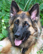
Actually … I didn’t want to really learn about this but I guess I will…. It’s also called Degenerative Lumbosacral Stenosis or Lumbosacral Instability. I think it’s like my sciatica. Anyway… here’s the scoop from online:
Cauda equina syndrome (degenerative lumbosacral stenosis) is caused by compression of the nerve roots (cauda equina) coursing through the lumbosacral spinal canal in the lower back. Nerve root entrapment and pressure ca
The most common symptom is progressive sharp pain. However, this syndrome can manifest itself in a number of ways. Intermittent lameness in one or both pelvic (rear) limbs or a stilted gait is a common initial sign. The patient may progressively have more difficulty rising from a prone position or may be unusually reluctant to leap. The dog may act suddenly painful or lame immediately after getting up or jumping. Strenuous activity may exacerbate the signs. Vocal expression of pain may vary from moans or whimpers when the dog tries to rise to sharp cries or howls when touched over the rear quarters or when making a wrong move during exercise. Eventually even the most pain tolerant individuals will react to the burning pain of the nerve root entrapment caused by this syndrome. Chewing at the tail or rear feet as well as bowel and bladder incontinence may be seen in advanced cases where severe pressure on the nerve roots causes a burning sensation. The most devastating cases can evolve to full paralysis.
Diagnosis
The neurologic examination begins by observing the gait. Specific tests for pain and neurologic dysfunction are then performed to confirm the site of the lesion.
Individuals with hip dysplasia will often show a mild response to hip extension whereas dogs with lumbosacral disease will object more acutely to hip extension and cry when pressure is added to the lumbosacral junction (see Fig.1). Manipulation and hyperextension of the tail cause
s an exquisite pain response. The spinal reflexes are tested, including the perineal reflex and anal tone, to assess the early signs of nerve root entrapment.
Nerve root entrapment and pressure can result from an arthritic process, infection, a degenerative disc rupture, or tumors. Therefore, it is essential to accurately diagnose the animal's problem before considering treatment (see Figs. 2 and 3). This requires radiography (x-rays).
Plain radiographs may not be useful in diagnosing such things as infection. A definitive diagnosis may require a myelogram or epidurogram (contrast dye studies of the spine) to confirm not only the location of the lesion but also the position of any ruptured discs in relation to entrapped nerve roots as the spine is flexed and extended. The myelogram and epidurogram are common and safe diagnostic procedures when performed under the proper conditions. In difficult cases, MRI or CT scans are of exceptional diagnostic value. Electromyography (EMG) may be of value in substantiating the diagnosis and the severity and symmetry of nerve root entrapment.
Treatment
Medical therapy consisting of rest and antiinflammatory/analgesic medications should be attempted in patients experiencing an initial episode with only mild pain.
Indications for surgical intervention include neurologic deficits, pain unresponsive to conservative treatment, and frequent recurrences of pain (even if the episodes respond well to medical treatment). To relieve pressure on the entrapped nerve roots, a dorsal laminectomy is performed. This involves removing portions of the bony spinal canal surrounding the entrapped nerve roots. The nerve roots (cauda equina) are then gently retracted to one side with blunt nerve hooks exposing any herniated discs as a large dome on the floor of the spinal canal. Any herniated discs are excised, compressive osteophytes are removed, and foramenotomies (opening the nerve root canals) are performed to relieve root entrapment. Once the pressure is relieved, neurologic function gradually returns.
Postoperative Care
A course of rest is the most important component of postoperative care. All strenuous activity should be curtailed for at least six weeks. At that time the exercise level is gradually increased. If the dog is obese, weight should be reduced.
The prognosis depends on the severity and chronicity of clinical signs before surgery. Dogs with pain, reluctance to jump, or tenderness upon getting up as their only symptoms will usually improve rapidly and dramatically. Some patients may have an occasional, transient, painful episode. Dogs with chronic neurologic dysfunction will take much longer to improve, and they may never return to completely normal function. However, at the very least they will return to a pain free lifestyle.


